Histopathology, expedited
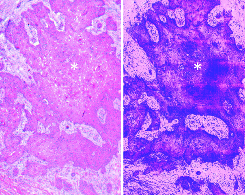
Our product is a novel imager that produces high-resolution, 3D digital pathology images of biopsied or surgically-removed tissue specimens at the point of care.
Why is this important?
Rapid, digitally colored histopathology images will enable physicians to make real-time clinical decisions, delivering significant time and cost savings for the clinic and an enhanced patient experience.

Mohs Surgery and Skin Cancer Patients
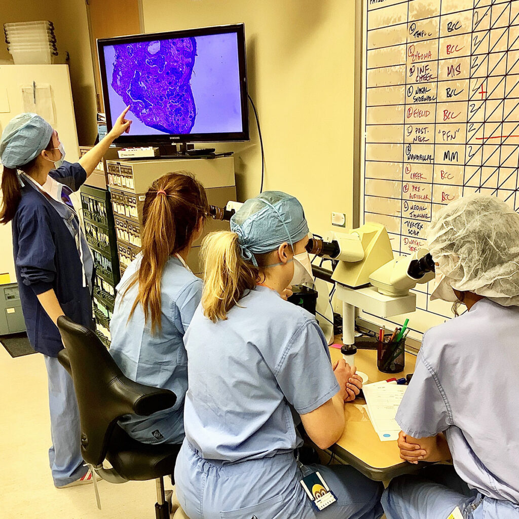
Mohs Surgery, a process of removing layers of cancer tissue for treatment of skin cancer, depends on standard histopathology for diagnosis, creating long and uncomfortable wait times for patients. SurgiVance’s advanced surgical pathology device aims to bring rapid diagnosis to the point of care and reduce Mohs surgery time from hours to minutes.